Organoids
Organoids are small, self-assembling three-dimensional multicellular structures that mimic organs and can be grown outside the body. Their ability to recapitulate the complexity of organ structures and mirror gene and protein expression makes organoids useful for various biomedical, research and drug development applications.
How does the cellular composition of organoids differ from 2D and other 3D cell cultures?
The most common type of cell culture is two-dimensional (2D) cell culture. 2D cell culture has proved to be a critical model for investigating the nature of cellular processes and molecular events. Researchers devised cell culture to grow cells under controlled conditions outside their physiological environment. Adherent cells are planar as they are grown in a monolayer on a solid support.
Where the 2D cell culture model falls down is that in vivo, the situation is very different. In vivo, cells and tissues are surrounded and supported by the extracellular matrix (ECM). The ECM consists of extracellular macromolecules (collagen, laminin, glycoproteins and proteoglycans), ECM-associated signalling molecules and connective tissue containing stromal cells. These components support and regulate a myriad of cell processes (morphology, differentiation, polarity and division). 2D cell culture lacks ECM and is grown on a rigid support. This, in turn, affects the composition and morphology of cells and the spatial arrangement of cell surface receptors.
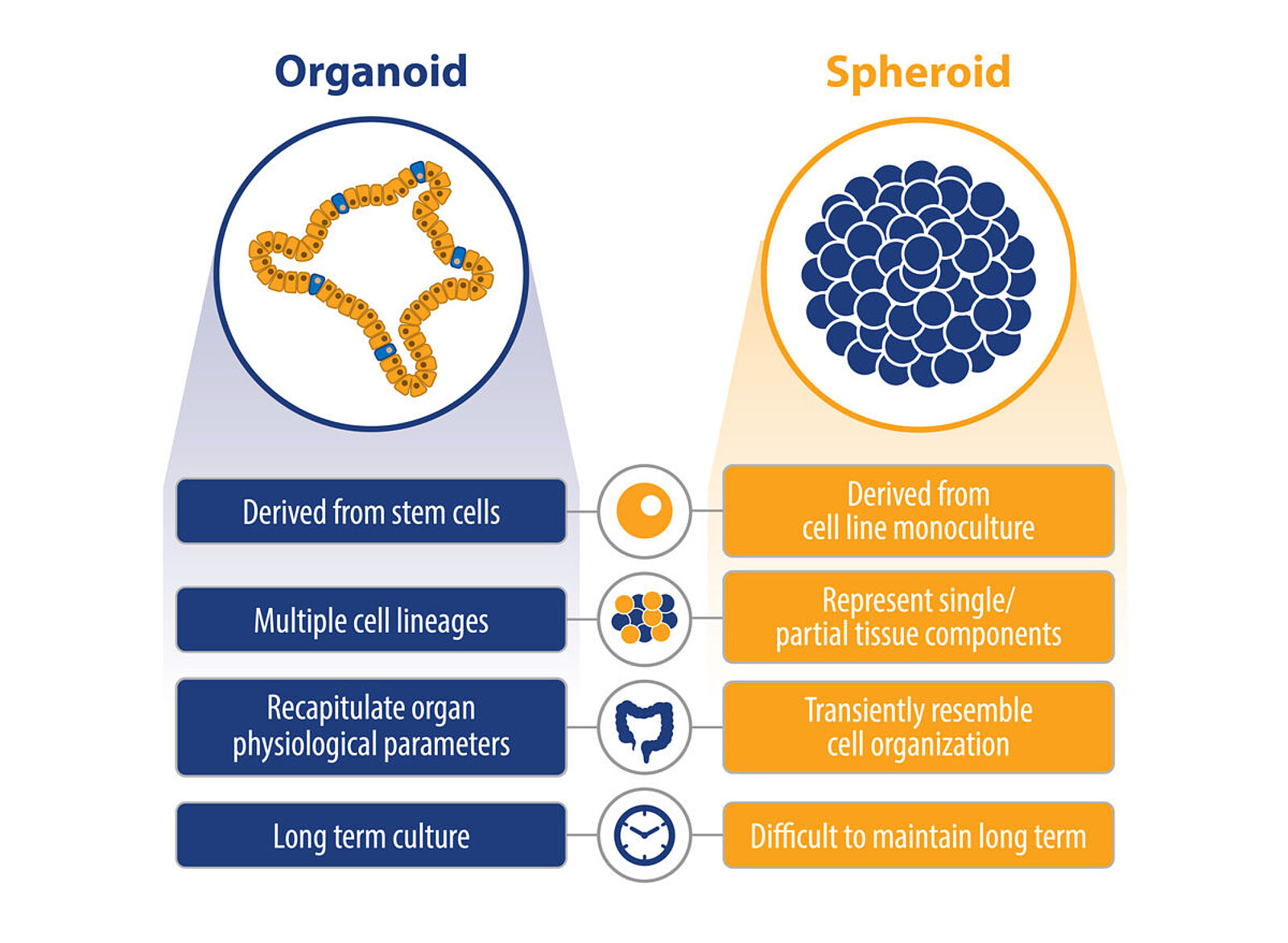
3D cell cultures are more physiologically accurate than 2D cell models as they more closely resemble the in vivo environment. 3D cultures allow cells to be grown in an ECM. Organoids are a type of 3D culture. Another type of 3D culture is spheroids. Spheroids are cell aggregates composed of a single cell type or several cells, which can be immortalised in cell lines, primary cells or human tissue.
Organoids contain either pluripotent stem cells (PSCs) or adult stem cells (AdSCs). AdSCs are multipotent cells that can give rise to multiple cell types and can be obtained from tissue biopsies. Cancer organoids are generated from cancer stem cells. AdSCs are more limited than PSCs, which can give rise to all cell types. Organoids can be generated with or without scaffolds, such as hydrogels.
First organoid model
The first organoid model was developed with the aim of getting a more physiological model to study the intestinal epithelium. Hans Clevers and teams at the Hubrecht Institute in the Netherlands first started culturing stem cells expressing the receptor, Lgr5, for Leucine-rich repeat-containing G-protein coupled receptor 5, which were derived from the crypt or base of the epithelium. It is known that Wnt signalling is crucial for crypt proliferation. Lgr5+ cells were selected as Lgr5 enhances the Wnt signalling pathway. The researchers devised a simple cocktail that excluded serum and used three recombinant proteins, EGF, Noggin, and R-spondin1 (RSPO1), all grown in a matrix using Matrigel.
R-spondin-1 (RSPO1) also acts as a Wnt agonist, increasing cell production in the crypt. EGF was responsible for the intestinal proliferation, and Noggin led to an expansion of crypt numbers (Sato et al., 2009). The Matrigel's essential role was to provide a typical tissue environment, as laminin is found at the base of the crypt.
The single stem cells (Lgr5) first formed a cyst structure but, once dissociated and replated, went on to form organoid structures. The Lgr5+ cells were multipotent stem cells capable of forming all the cell types of the gut epithelium. This resulted in the development of a mini-version of the gut epithelium, which then paved the way for the long-term culture of other organoid models.

Applications of organoids
Organoids have been influential in developmental biology and have critical applications in the preclinical testing phase in drug discovery, personalised medicine and regenerative medicine.
In the past, 2D cell culture has been commonly used for drug discovery. The drawback of this technique is that drug candidates that have proven successful in 2D cell culture models have gone on to fail in later-stage testing. 2D cell culture produces different responses to 3D culture, which closely mimics the physiological environment. The reasons behind this may be the differences in morphology and the expression of cell surface receptors, often targeted by pharmaceutical compounds (reviewed in Jensen & Teng, 2020).
Organoids provide a significant biological advantage over 2D cell culture because they resemble the tissue microenvironment. They display a more biologically relevant response to pharmaceutical compounds and are also alternatives to animal testing. Animals are widely used in the pharmaceutical industry to predict human toxicity, which dates back to the 1930s. However, animal models have been shown to be poor predictors of drug safety in humans (van Norman, 2019). This is why the recent amendment — FDA Modernization Act 2.0 — called the "Animal Testing Alternatives" will now allow other substitutes for animal testing, such as organoids, to assess the safety and effectiveness of a drug.

LubioScience provide essential components for organoid culture
LubioScience work with MBL International ect. to provide reagents for organoid culture.
Culturing organoids requires cell culturing conditions and equipment, as organoids are derived from stem cells capable of growth and expansion over time. LubioScience provide serum-free media containing Wnt3a and Afamin. As Wnt3a is fat-soluble, it forms aggregates in serum-free media, preventing it from exerting its activity. Afamin is a member of the albumin family of proteins present in serum and acts to stabilise Wnt3a when complexed together, making long-term culture of organoids using Wnt 3a possible.
As highlighted in the development of the first intestinal organoid structures, growth factors and cytokines are also important for culturing organoids. LubioScience provide a range of recombinant growth factors and cytokines, including EGF, Noggin (NOG), R-spondin (RSPO1), and BMP.
Please visit our website for more information on conditioned media, recombinant growth factors and cytokines essential for organoid culture. LubioScience are happy to support you with your application needs.
Bethyl Laboratories - Fortis Life Sciences applies B-cell sorting and recombinant DNA technology to deliver high quality recombinant rabbit monoclonal antibodies. The pillar strategy validation ensures that the antibody works in the stated applications.
ACROBiosystems
ACROBiosystems is a leading manufacturer of recombinant proteins and other critical reagents to support the development of target therapeutics, vaccines, and diagnostics.
MCE - MedChemExpress
MedChemExpress offers high quality small molecules, reference compounds, APIs & natural compounds and inhibitor libraries. Supreme quality and rigorous QC testing by HNMR, LC-MS and HPLC - stability testing. Most compounds are tested for activity.
TargetMol
TargetMol offers compound libraries and a wide range of high-quality inhibitors, activators, natural compounds, peptides, antibodies, and novel life-science kits for laboratory and scientific use.
References
- Jensen, C., & Teng, Y. (2020). Is It Time to Start Transitioning From 2D to 3D Cell Culture? In Frontiers in Molecular Biosciences (Vol. 7). doi.org/10.3389/fmolb.2020.00033
- Sato, T., Vries, R. G., Snippert, H. J., Van De Wetering, M., Barker, N., Stange, D. E., Van Es, J. H., Abo, A., Kujala, P., Peters, P. J., & Clevers, H. (2009). Single Lgr5 stem cells build crypt-villus structures in vitro without a mesenchymal niche. Nature, 459(7244), 262–265. doi.org/10.1038/nature07935
- van Norman, G. A. (2019). Limitations of Animal Studies for Predicting Toxicity in Clinical Trials: Is it Time to Rethink Our Current Approach? JACC: Basic to Translational Science, 4 (7). https://doi.org/10.1016/j.jacbts.2019.10.008
Images
- Figure 1. https://blog.crownbio.com/organoids-or-spheroids
- Figure 2. Kriz, V.; Korinek, V. Wnt, RSPO and Hippo Signalling in the Intestine and Intestinal Stem Cells. Genes 2018, 9, 20. https://doi.org/10.3390/genes9010020