Synaptic Systems strives to provide you with the best antibodies and camelid single domain FluoTags for neuroscience and cell-biology. Our in house developed products are validated for a broad panel of applications like westernblot (WB), immunocytochemistry (ICC), immunohistochemistry (IHC, IHC-P), immunoprecipitation (IP) and ELISA to guarantee the best quality and reliability.
Products
- Antibodies for neuroscience
- Antibodies for cell biology
- FluoTags - camelid single domain antibodies
- HistoSure - antibodies for histopathology
- Resins, chemicals, kits & lysates
- Secondary antibodies
HistoSure anti CD11b antibody for histopathology
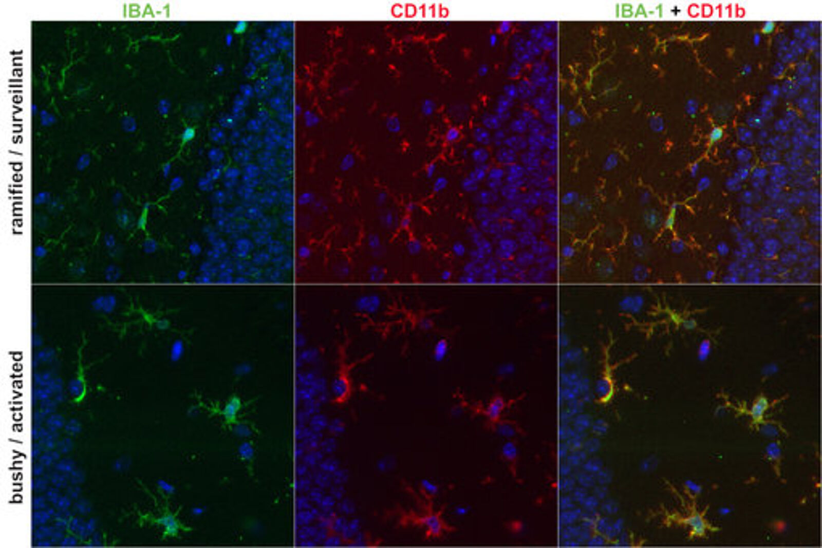
Microglia are the key immune effector cells of the central nervous system (CNS), including the brain, spinal cord, retina and olfactory bulb. Microglia function in the support of CNS development, in sustaining homeostasis and in immune response. Surveying or ‘resting’ microglia are ramified and build a dense network spanning the CNS.
Microglia activation leads to increased expression of IBA-1, MHCII, CD68 and CD11b, also known as integrin alpha M (ITGAM). CD11b expression in microglia is induced in response to Nitric Oxide (NO) produced by the inducible nitric-oxide synthase iNOS. Together with CD18, CD11b forms the integrin complement receptor 3 (CR3), which is involved in adhesion processes, phagocytic elimination of pathogens, induction of both inflammatory and tolerogenic responses, and modulation of parallel or downstream host defense pathways.
The image on the right shows PFA fixed brain sections from a wild-type mouse (control brain; upper row) and a sepsis mouse model (PCI brain; lower row). Sections were immunostained with rat anti-CD11b (cat. no. HS-384 117, red channel) and Guinea pig anti-IBA1 (cat. no. HS-234 004, green channel). Nuclei were visualized by DAPI staining (blue channel).
FluoTag - single domain antibodies
FluoTags are camelid single domain antibodies consisting only of one antigen binding site of a Alpaca heavy chain antibody. With only ~15 kDa, these Tags are about 10-times smaller than conventional IgG antibody molecules.
Two variants of FluoTags are available, the -Q and -X series:
In FluoTag-Q each fluorophore is coupled to exactly one FluoTag, which in turn binds to its target molecule in a monovalent fashion. The high binding affinity and a coupling efficiency of > 95% guarantees a highly linear relation between the number of target molecules and the intensity of fluorescence. This enables a direct count of the target molecule of interest. The fluorophore is located exceptionally close to the recognized epitope (less than 1.5 nm), which is ideal for all microscopy techniques.
In FluoTag-X two fluorophore molecules are site-specifically coupled to each FluoTag molecule. Therefore, the reagent simultaneously targets up to four fluorophores to the protein of interest, which ensures extra-bright signals. Owing to the small size of the FluoTags, the distance between the target epitope and each fluorophore is below 4 nm.
In comparison to detection systems using conventional antibodies, FluoTag-X can thus improve the localization accuracy by 10-15 nm. Both features - superior brightness and precise fluorophore placement - render the FluoTag-X products excellent tools for all microscopy techniques.
