Biotium is a leading life science reagent manufacturer and supplier devoted to providing high-quality and innovative fluorescent tools that fuel scientific discovery.
Biotium is uniquely poised to invent cutting-edge products through the synergy of chemistry and biology. Our collaborative team of experienced chemists and biologists, who are at the forefront of fluorescent dye design, apply chemistry-based principles toward producing solutions for unmet challenges in life science and medical research. Ultimately, we aim to be a powerful force for accelerating groundbreaking research that results in significant contributions to overall scientific knowledge and human health.
Portfolio
- PCR & DNA Amplification
- Antibodies, Conjugates, & Labeling
- Antibody & Protein Labeling Kits
- Cellular Stains & Assays
- Protein Detection & Analysis
- Reagents & Accessories
ExoBrite™ CTB EV Staining Kits
Technology Highlight: ExoBrite™ CTB EV Staining Kits
EV staining you can trust
ExoBrite™ CTB EV Staining Kits are designed for robust staining of isolated EVs by flow cytometry or microscopy with minimal background.
Features
- Bright and specific staining of EV and exosome membranes
- Suitable for both purified and bead-bound exosomes
- Optimized for detection by flow cytometry
- Compatible with antibody co-staining
ViaFluor® SE Cell Proliferation Kits
Cell Proliferation Monitoring by Flow Cytometry
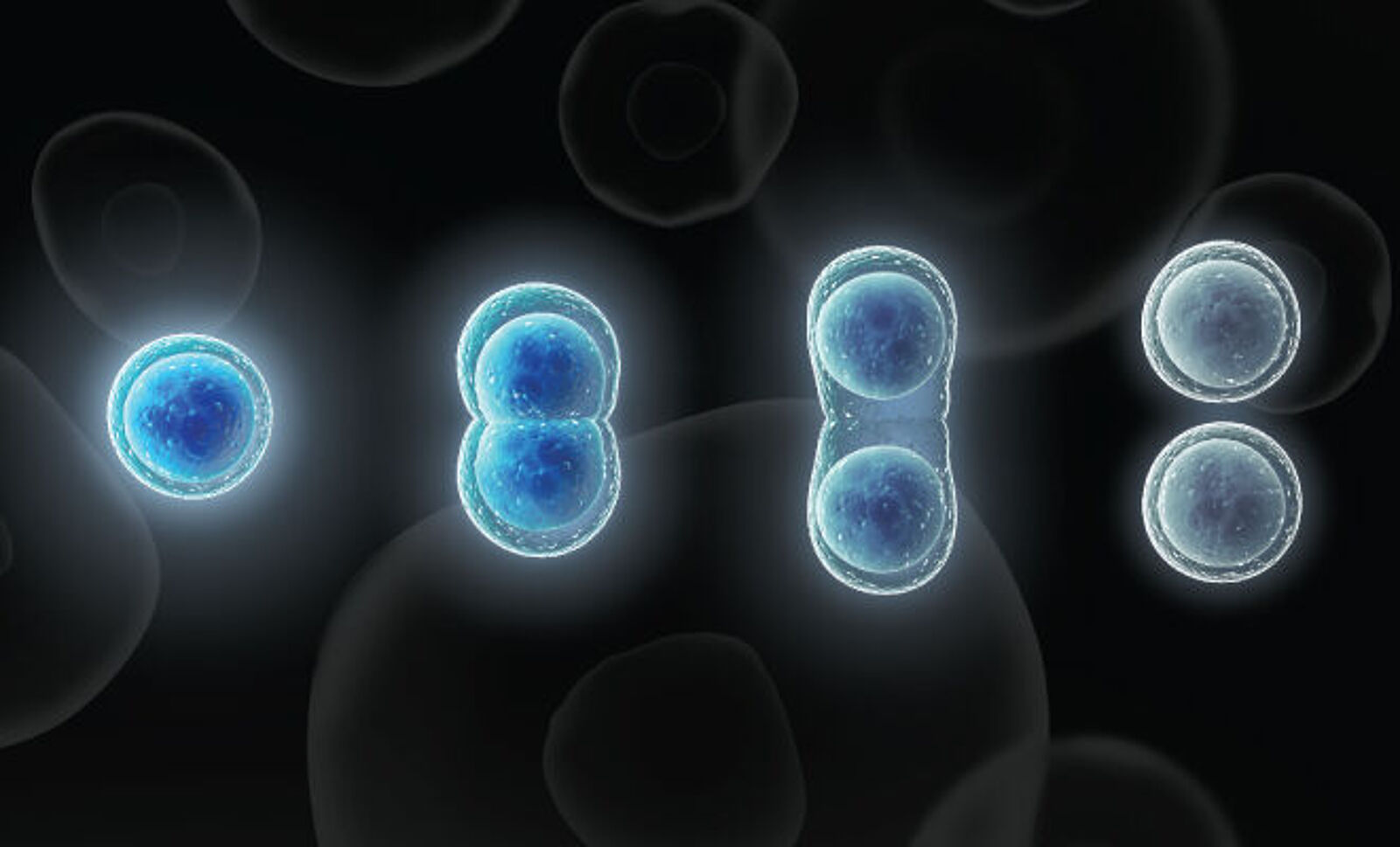
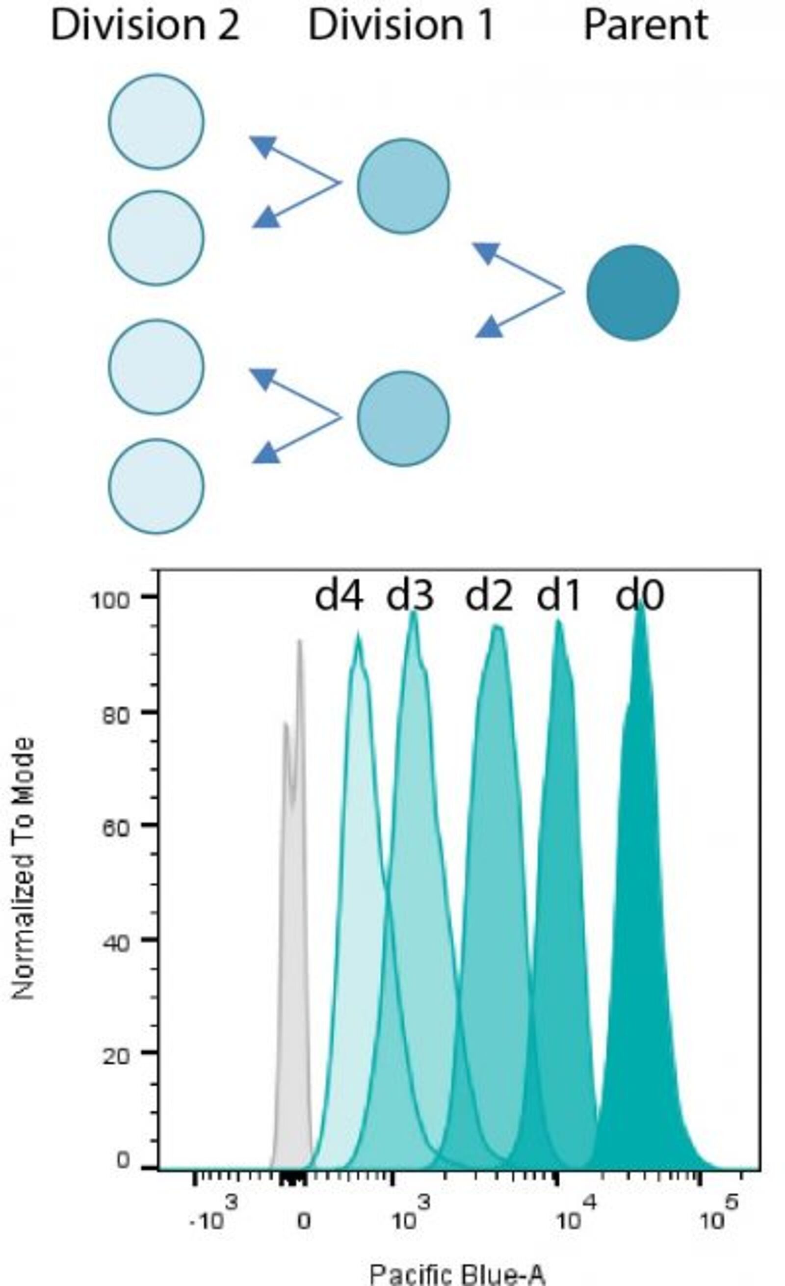
Cell proliferation dyes diffuse passively into live cells and are used for long-term cell labeling. They are initially non-fluorescent esters, but are converted to fluorescent dyes by intracellular esterases and will covalently react with amine groups on intracellular proteins at the same time, forming fluorescent conjugates that are retained in the cell. Immediately after staining, a single bright fluorescent population will be detected by flow cytometry. Each cell division that occurs after labeling is revealed by the appearance of a successively dimmer fluorescent peak on a flow cytometry histogram (Figs. 1&2). Cell proliferation dyes can be used to track cell divisions in vivo or in vitro. The staining can withstand fixation and permeabilization for subsequent immunostaining.
ViaFluor® SE Cell Proliferation Kits
CFSE (also known as CFDA-SE or carboxyfluorescein diacetate, succinimidyl ester) has been used for many years to monitor cell proliferation by flow cytometry. While several alternatives have been developed in more recent years, CFSE remains popular and widely used. Biotium continues to offer CFSE under the tradename ViaFluor® CFSE. However, CFSE has several drawbacks including leakage from the cell, cell toxicity, and bleed-through into the PE and PE-TexasRed® channels. ViaFluor® 405 SE and ViaFluor® 488 SE cell proliferation dyes were developed at Biotium to provide superior cell staining, fixability, and low toxicity. ViaFluor® 405 is excited with the violet laser and detected in the Pacific Blue® channel, and gives great peaks with no toxicity (Fig. 1). ViaFluor® 488 and ViaFluor® CFSE are both detected in the FITC channel, but ViaFluor® 488 was developed as a less toxic, less leaky and more fixable alternative to the classic dye CFSE. It also has less bleed-through into other channels such as PE and PE-Texas-Red®.
Methylene Blue Derivatives
Methylene Blue (MB) is a commonly used redox indicator in nucleic acid research. It is also studied for its use in medical applications as well as being used as a general biological stain. Reactive formats of MB can be conjugated to biomolecules. The conjugate will have a blue color and be able to complex with nucleic acids.
Cholera Toxin Subunit
Cholera toxin is the symptom-causing toxin produced by the bacteria Vibrio cholerae during cholera infection. The toxin is composed of two subunits, A and B. Subunit A is the toxic enzymatic subunit present in one copy per toxin. CT-B is the receptor binding subunit that is found as a pentamer in each toxin and is relatively non-toxic, making it useful for cell biological studies. CT-B has been used as a neuronal tracer and has also been shown to bind to GM1 gangliosides that are found in lipid rafts on the surface of mammalian cells. Therefore, fluorescently labeled conjugates of CT-B have been used as lipid raft markers and endocytic tracers for live imaging or on fixed cells.
Superior CF® Dyes
Biotium’s next-generation CF® Dyes were designed to be highly water-soluble with advantages in brightness and photostability compared to other commercially available dyes. Learn more about CF® Dyes.
Note: Conjugates of blue-fluorescent dyes like CF®350, CF®405S and CF®405M are not recommended for detecting low abundance targets and may be challenging to use in tissue specimens. Blue dyes have lower fluorescence and photostability, and cells and tissue have high autofluorescence in blue wavelengths, resulting in lower signal to noise compared to other colors.