BPS Bioscience’s purpose is to advance new scientific discoveries that lead to therapies by creating innovative solutions for research. BPS Bioscience are experts in protein design, expression, purification, and characterization, cell line and lentivirus engineering, and biochemical and cellular assay development. Their products and services have enabled thousands of researchers to advance biological discoveries across a wide variety of disciplines, including epigenetics, cell signaling, SARS-CoV-2, immunotherapy, adoptive cell therapies, and many more.
Portfolio
- Cell lines, including reporter cell lines
- Biochemical Assay Kits (Luciferase Assays, PARPtrap™ Assay, ...)
- Proteins
- Primary Cells
- Cell Media and Luciferase Reagents
- AAV, Lentiviruses, VSV
- Cell-Based Assay Kits
- Antibodies
- Inhibitors/Activators
- Cell Isolation Kits
BPS Bioscience focus on emerging research needs and rapidly deliver tools to enable progress. When new research projects face limited assay solutions, they can quickly develop novel products and services that are effective and consistent.
- Catalogue - BPS Bioscience’s ever-expanding catalogue of over 4500 products has enabled discoveries worldwide from small academic labs to large pharmaceutical companiess. Thier products have been extensively cited with increasing numbers each year. BPS Bioscience understands that science never stands still, so they strive to stay at the forefront of discovery. Every month, they release dozens of useful, high-quality products.
- Quality - BPS Biosceince focus on developing quality products, providing excellent customer service, and maintaining a robust quality management system. They are ISO 9001:2015-certified, ensuring constant monitoring and continuous improvement in order to meet customer needs.
Assay kits
Luciferase Assay Systems
BPS Bioscience's luciferase assay systems (both ONE-Step and TWO-Step Luciferase (Firefly & Renilla) Assay System) are sensitive, stable, and convenient reagents to use for quantification of luciferase signals.
ONE-Step & TWO-Step Luciferase Assay Systems
BPS Bioscience's luciferase assay systems (both ONE-Step and TWO-Step Luciferase (Firefly & Renilla) Assay System) are sensitive, stable, and convenient reagents to use for quantification of luciferase signals.
- ONE-Step Luciferase Assay System: The ONE-Step Luciferase Assay System is designed to be used for high-throughput, sensitive quantitation of firefly luciferase activity in mammalian cell culture.
- TWO-Step Luciferase (Firefly & Renilla) Assay System The TWO-Step Luciferase (Firefly & Renilla) Assay System is designed to be used for high-throughput, rapid quantitation of both Firefly and Renilla luciferases from a single sample in mammalian cell culture.

PARPtrap™ Assay Kit for PARP1
Description:
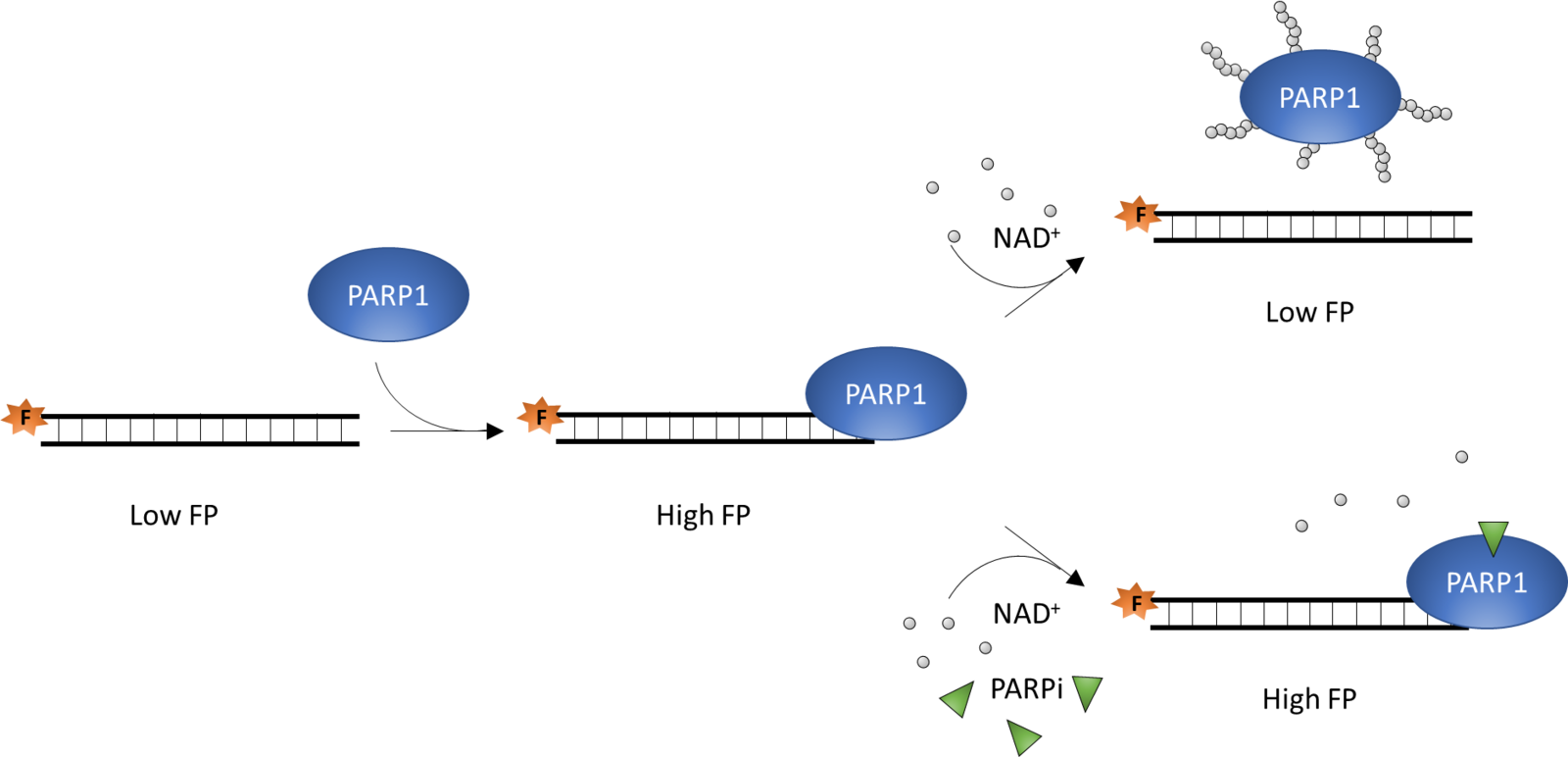
The PARPtrap™ Assay Kit for PARP1 is designed to measure PARP1/DNA complex formation in a high throughput screening assay using fluorescence polarization (FP). PARP1 is known to bind damaged DNA through its DNA-binding domains. Binding to DNA activates PARP1 and in the presence of NAD+ PARP1 ribosylates itself (auto-ribosylation), what in consequence leads to PARP1 dissociation from the DNA due to the accumulated negative charge of the ribosyl polymer. In the presence of some inhibitors, however, PARP remains bound to the DNA, a phenomenon termed trapping. Trapped PARP-DNA complexes have been shown to be highly cytotoxic to cancer cells, therefore such inhibitors may be desirable for cancer treatment.
The key to the PARPtrap™ Assay Kit for PARP1 is the fluorescent-labeled oligonucleotide duplex. In the absence of ribosylation, PARP1 binds to the fluorescent probe, forming a large complex and resulting in the emission of highly polarized light. However, after auto-ribosylation, PARP1 dissociates from the oligonucleotide duplex, which then rotates freely, emitting less polarized light (Figure 1). Addition of a PARP1 inhibitor results in trapping of PARP1 to the fluorescent oligonucleotide duplex, and increases the FP signal in a dose dependent manner.
The PARPtrap™ Assay Kit for PARP1 is a fluorescence polarization homogeneous assay. The FP signal is measured using a fluorescent microplate reader capable of measuring fluorescence polarization.
Cell lines
BPS Bioscience has developed over 250 stable cell lines applicable across a wide range of research areas including immunotherapy, CAR-T cell therapy, cell signaling, metabolism, neuroscience, and much more. We now also feature iPSC-derived cells for drug discovery.
More about BPS Bioscience stable cell lines
Top sellers
TL1A-Responsive Luciferase Reporter Jurkat Cell Line
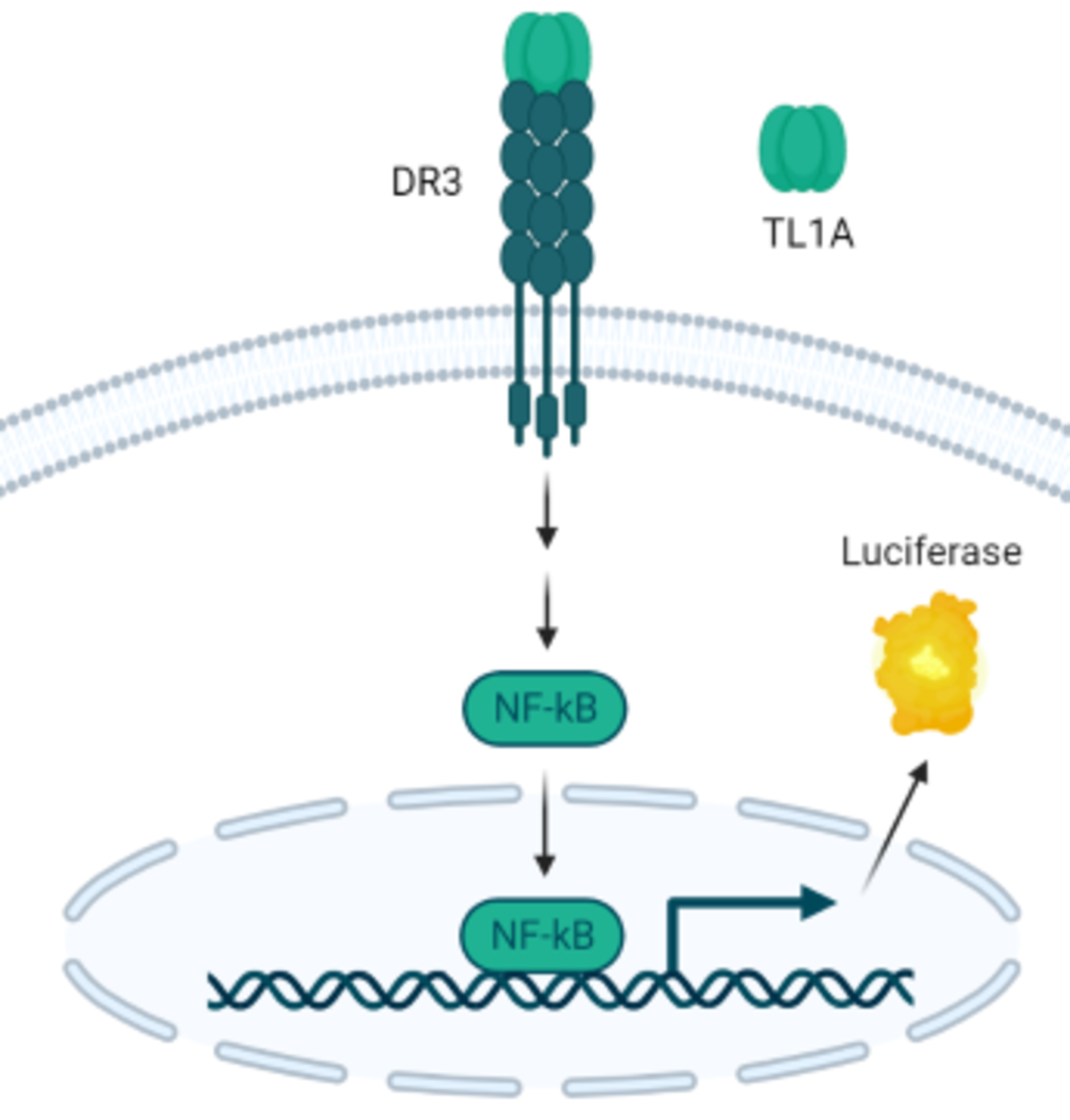
TNF-like ligand 1A (TL1A, also known as Vascular endothelial growth inhibitor, VEGI or TNFSF15) is an anti-angiogenic cytokine. It is an important mediator of inflammation, participates in innate and adaptive immune homeostasis through binding to its receptor, DR3, activating downstream signaling. Numerous studies showed that soluble TL1A can be detected in the serum of patients with T-cell mediated autoimmune diseases like rheumatoid arthritis, psoriatic arthritis, and inflammatory bowel disease. In addition, recent clinical studies suggested that anti-TL1A antibody treatment is a promising therapeutic approach in inflammatory and auto-immune disorders.
Description:
TNF-like ligand 1A (TL1A)-Responsive Luciferase Reporter Jurkat Cell Line is a TL1A-responsive DR3/NF-kB luciferase reporter Jurkat cell line expressing firefly luciferase under the control of an NF-kB response element, and with stable expression of human DR3 (death receptor 3; TNFRSF25; NM_003790.3). Expression of the firefly luciferase gene is driven by NF-kB response elements located upstream of the minimal TATA promoter. Activation of the NF-kB signaling pathway by the DR3 ligand TL1A (TNF-like Protein 1A; TNFSF15) can be monitored by measuring luciferase activity.
GLP-1R/CRE Luciferase Reporter HEK293 Cell Line
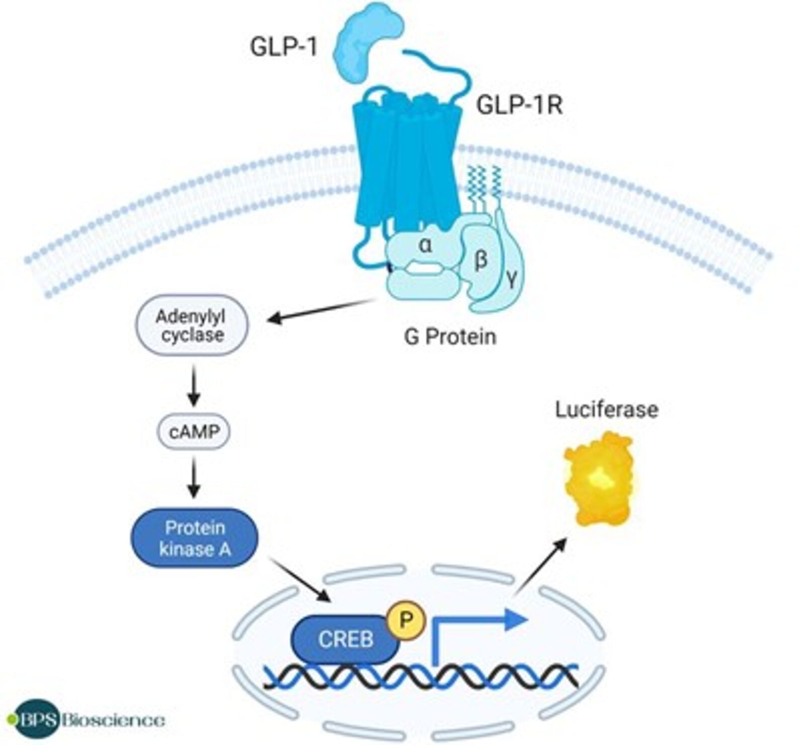
GLP-1R, a member of the class B family of G protein-coupled receptors (GPCRs or secretin-like receptors), is a transmembrane protein primarily found in pancreatic β cells and brain neurons and is activated by the peptide hormone glucagon-like peptide 1 (GLP-1) and glucagon that is secreted from intestinal L-cells after nutrient ingestion. GLP-1R plays an important role in controlling blood sugar level by enhancing glucose-stimulated insulin secretion, glucose, lipid metabolism, and satiety. Its role in the brain seems to be in the control of appetite.
Research efforts have focused on the regulation of the GLP-1R mediated signaling pathway as a therapeutic approach to type 2 diabetes (T2DM) and have resulted in the development of several GLP-1 FDA-approved agonists. In addition to their role in insulin secretion, GLP-1R agonists can also contribute to weight management, decrease the potential for cardiovascular diseases and protect beta cells. A role in tumor development in patients with T2DM is also being investigated but further studies are required to fully understand the functions of GLP-1R and its agonists.
Description:
GLP-1R/CRE Luciferase Reporter HEK293 Cell Line is an engineered HEK293 cell line expressing firefly luciferase under the control of cAMP response element (CRE), and human GLP-1R (Glucagon-like peptide 1 receptor; accession number BC113493). Activation of GLP-1R in these cells can be monitored by measuring luciferase activity.
The functionality of GLP-1R/CRE Luciferase Reporter HEK293 Cell Line was validated in dose-response assays using the peptide agonists Glucagon-like peptide 1 (7-37), Exendin-4, Lixisenatide, GLP-1 moiety from dulaglutide, Semaglutide, Tirzepatide and Retatrutide. This cell line has also been validated in a dose-response assay using a small molecule agonist, Danuglipron. GLP-1R/CRE Luciferase Reporter HEK293 Cell Line responds to agonists by inducing luciferase activity in a dose-dependent manner.
OX40/NF-κB Luciferase Reporter HEK293 Cell Line
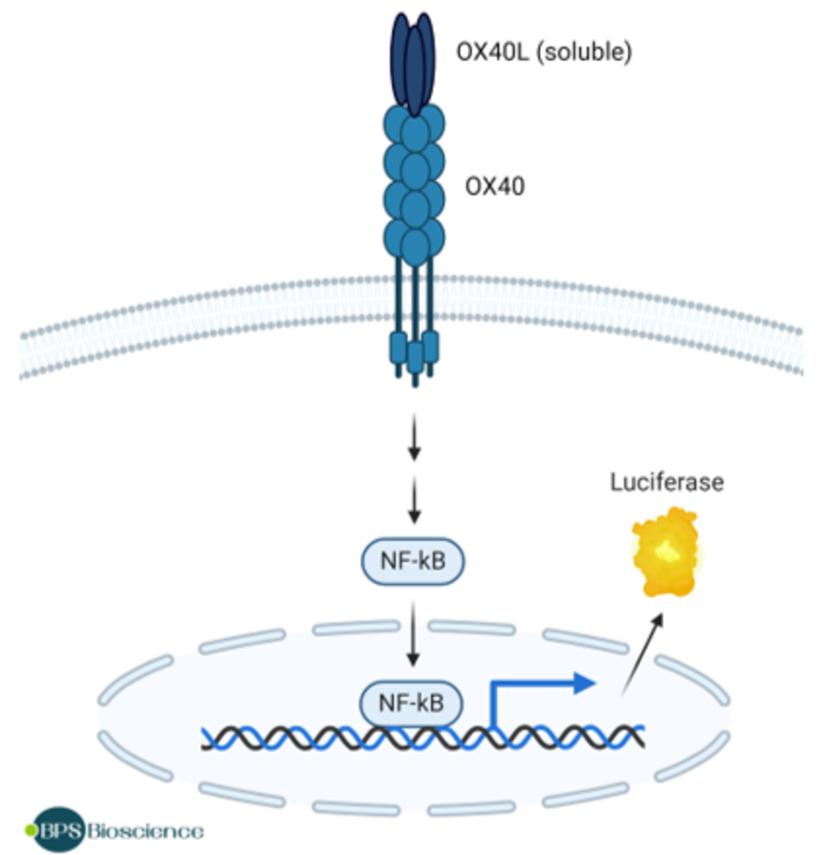
OX40, also known as CD134, is a co-stimulatory receptor, of the TNF (tumor necrosis factor) receptor family, expressed on the surface of T cells. Binding of OX40 to its ligand, OX40L (also known as CD252), potentiates T cell activation, differentiation, proliferation, survival and T cell effector function. OX40L is present in NK cells, participating in their activation and cytotoxicity profile, and dendritic cells. OX40 can bind to members of the TRAF (TNFR associated factor) family of proteins, which can then regulate the NF-κF (nuclear factor kappa-light chain enhancer of activated B cells) signaling pathway. OX40 and OX40L can be found in cancer cells, such as AML (acute myeloid leukemia) and breast cancer cells. Studies have shown that OX40 agonists can increase anti-tumor immunity and improve tumor-free survival in pre-clinical studies. Alternatively, OX40 antagonists offer potential as therapeutics for inflammatory diseases. The development of new modulators of the OX40/OX40L activity are promising therapies for patients suffering from solid tumors or auto-immune disorders.
Description:
The OX40/NF-κB Luciferase Reporter HEK293 Cell Line is a HEK293 cell line expressing the Firefly luciferase reporter under the control of NF-κB (nuclear factor kappa light chain enhancer of activated B cells)-responsive elements and OX-40 (Tumor Necrosis Factor Receptor Superfamily Member 4, GenBank Accession No. NM_003327).
This cell line has been validated by flow cytometry, and with OX40L and anti-OX40 antagonist antibodies.
Limited use license for BPS cell lines
Please note that purchase of cell lines from BPS requires a limited use license for non-commercial use. You can download the license agreement below. The limited use license must be completed and signed by the customer prior to purchasing any cell lines from BPS.
| Filename | Download |
|---|---|
| BPS Bioscience Limited Use License Agreement |
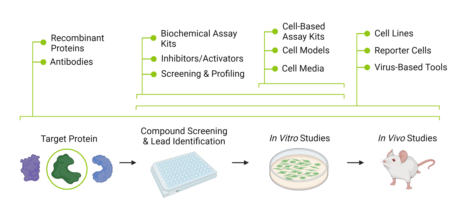
How BPS Bioscience Products Drive Pre-Clinical Drug Discovery
BPS Bioscience products are intended to enable research through each phase of the pre-clinical drug development process. This infographic outlines where our different product types can be used. Some can be used across many research phases. For example, cell lines can be during the compound screening phase to analyze toxicity, while fluorescent cell lines could be used for in vivo studies. Our collection of complementary products provide a suite of solutions that ultimately drive research throughout early drug discovery.
Contact form for BPS Bioscience cell lines
Please don't hesitate to reach out to our team to request a quote or for any questions. We're here to help.
You can find more information in our Privacy policy