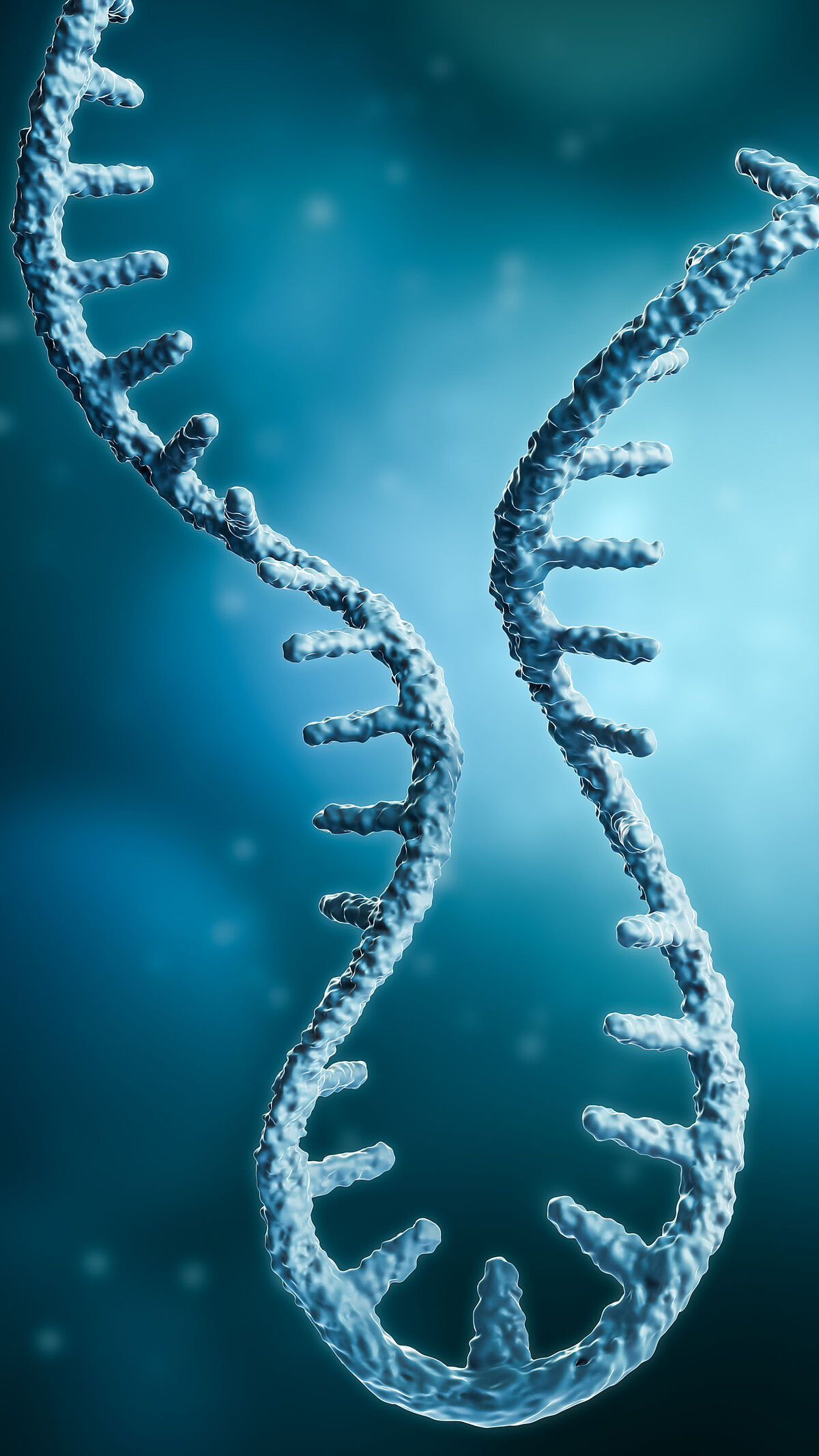
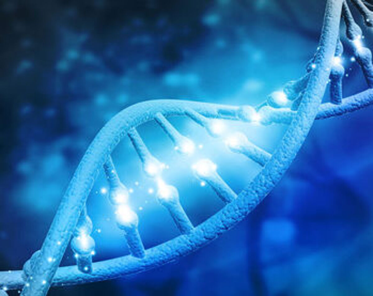
What is transcriptomics?
In multicellular organisms, not every gene is transcriptionally active. Which genes are switched on or off depends on cell type, developmental stage and physiological signals. Transcriptomics is the study of the complete set of ribonucleic acid (RNA) molecules – coding and non-coding – expressed in a given cell, tissue or organism. By examining transcriptomes, researchers can uncover how patterns of gene expression shape development and disease.
Early methods in transcriptomics
Before transcriptomics became established, messenger RNA (mRNA) transcripts were collected and converted into complementary DNA (cDNA) using reverse transcriptase in the late 1970s. Downstream analysis involved constructing cDNA libraries, followed by characterisation of cDNA clones with restriction enzymes, Southern blotting and hybridisation¹. By 1977, RNA transcripts could also be examined using Northern blots².
During the 1980s, Sanger sequencing was applied to random transcripts to generate expressed sequence tags (ESTs). Based on the incorporation of chain-terminating dideoxynucleotides, this lower-throughput method remained popular until higher-throughput approaches took over. In the late 1980s, reverse transcription was combined with PCR (RT-PCR or RT-qPCR), enabling RNA quantification³.
Transcriptomics was often paired with molecular cytogenetic techniques such as in situ hybridisation (ISH), which provided insights into the spatial and temporal distribution of RNA. These methods, however, were labour-intensive and limited to subsets of the transcriptome. In 1995, the first transcriptomic technique – serial analysis of gene expression (SAGE) – was introduced. Using Sanger sequencing of concatenated transcript fragments, SAGE enabled simultaneous quantitative analysis of large numbers of transcripts⁴.
The rise of microarrays and RNA sequencing
Two methods dominate modern transcriptomics: microarrays and RNA sequencing (RNA-Seq).
Microarrays, first described in 1995, detect transcript abundance by hybridisation to probes immobilised on glass slides. Extracted mRNA is converted into double-stranded cDNA, fragmented, and fluorescently labelled with Cy3 (green) or Cy5 (red). Hybridisation to probe arrays allows relative expression analysis of thousands of genes across two samples. While microarrays remain cost-effective and suitable for large studies, their use has declined since 2014⁵, largely due to the advent of RNA-Seq.
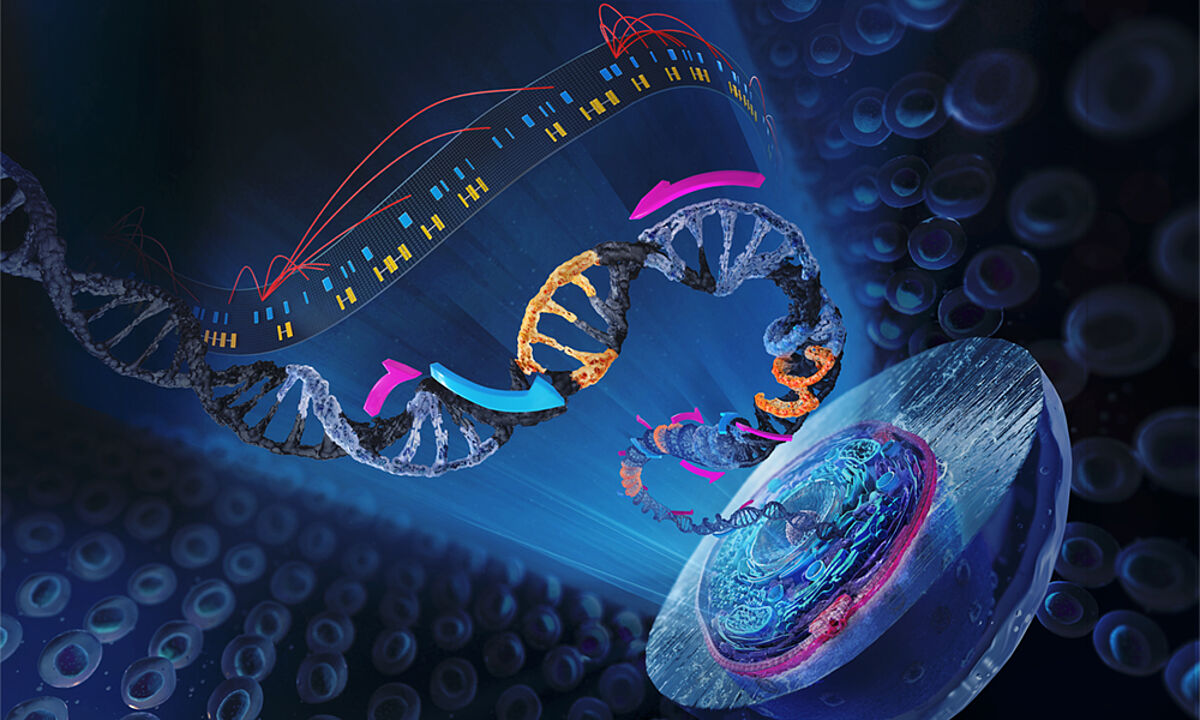
RNA-Seq, introduced in 2006 by Bainbridge et al.⁶, employs next-generation sequencing (NGS) to capture all RNA sequences in a sample. The workflow involves RNA extraction, purification, library preparation, sequencing, and data analysis. Unlike microarrays, RNA-Seq does not require prior sequence knowledge and can detect splice variants, single-nucleotide polymorphisms (SNPs) and other RNA species⁷. With sequencing costs dropping by over 90% between 2014 and 2020⁷, RNA-Seq has become increasingly accessible and widely adopted.
Comparing microarrays and RNA-Seq
| Characteristic | Microarray | RNA-Seq |
|---|---|---|
| RNA input | ~ µg quantities | ~ ng quantities |
| Prior knowledge | Required (reference transcripts for probe design) | Not required (genome sequence useful) |
| Sensitivity | ~10⁻³ (limited by fluorescence detection) | ~10⁻⁶ (limited by sequence coverage) |
| Sequence resolution | Requires dedicated arrays to detect splice variants | Detects SNPs and splice variants |
| Dynamic range | 10³ – 10⁴ (limited by fluorescence saturation) | >10⁵ (limited by sequence coverage) |
| Specificity | Cross-hybridisation can create signal noise | Free from probe redundancy issues |
| Data analysis | Standard lab software; minimal storage needs | Specialist bioinformatics tools; higher storage demands |
| Cost | Lower per sample for large studies | Cost per Gb decreasing |
Why spatial transcriptomics matters
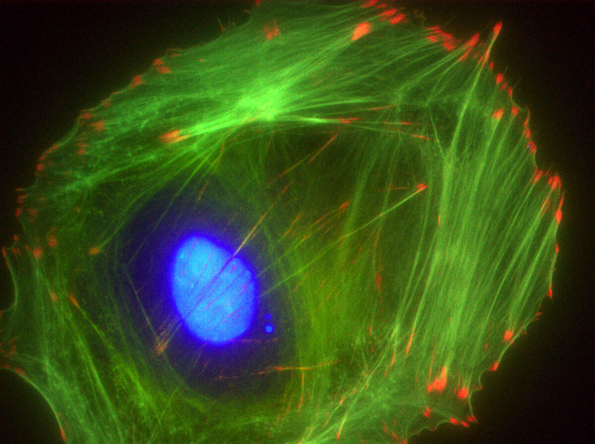
RNA-Seq is usually applied to bulk samples, such as pooled cells or tissue sections. While this provides valuable average gene expression data and highlights pathway regulation, it cannot resolve cell-to-cell differences. Single-cell RNA sequencing (scRNA-seq) addresses this by profiling individual cells and identifying rare populations hidden in bulk analyses.
However, scRNA-seq lacks spatial context. The cellular microenvironment plays a crucial role in regulating biological functions. For example, cancer stem cells (CSCs) depend on specific niches that maintain their properties⁸. On the mRNA level, evidence shows that transcript localisation affects gene function by influencing where proteins are produced and trafficked⁹.
Spatial transcriptomics (ST) overcomes this limitation by mapping gene expression within its native tissue environment. In recognition of its impact, Nature named ST the “Method of the Year” in 2020.
Spatial transcriptomics technologies
ST preserves the in situ location of transcripts while profiling gene expression¹⁰. The choice of method depends on desired throughput, resolution, sensitivity, sample type and instrumentation¹¹,¹². Broadly, ST methods fall into four categories:
1. In situ hybridization (ISH)
ISH visualises RNA directly within tissue sections. Fluorescent probes hybridise to complementary RNA sequences, generating signals that can be amplified and imaged. Sequential FISH (seqFISH), merFISH and seqFISH+ detect RNA molecules in single cells through repeated rounds of hybridisation, imaging and probe removal. Each transcript receives a barcode, allowing quantification and localisation. Originally developed for single cells, seqFISH has since been applied to complex tissues such as the mouse hippocampus¹³.
2. In situ sequencing (ISS)
ISS targets RNA in preserved tissues using padlock probes, which hybridise to specific transcripts and form circular templates. After reverse transcription, ligation and rolling circle amplification, fluorescent signals reveal transcript identity. Both “no-gap” and “gap-filling” probe approaches exist, offering sensitivity or direct sequence capture, respectively¹⁴. Variants include FISSEQ and Barista-seq, the latter employing Illumina sequencing for improved signal-to-noise ratios¹⁵.
3. In situ capture
Here, mRNA is extracted while retaining spatial information. Tissue sections are placed on glass slides coated with spatially barcoded primers. During permeabilisation, mRNA hybridises to these primers and is reverse transcribed into cDNA. Sequencing then maps transcripts back to their original tissue coordinates. This approach has been used in studies of the mouse brain and human breast cancer¹⁶, with newer adaptations enabling analysis of frozen and FFPE samples¹⁷,¹⁸.
4. Microdissection
Laser capture microdissection (LCM) isolates specific cells or regions from tissue under microscopic guidance. The extracted RNA is then sequenced, preserving native spatial information. Geo-seq, described in 2017, combines LCM with RNA-Seq to profile transcriptomes of dissected tissue regions¹⁹.
References
Sim, G. K., Kafatos, F. C., Jones, C. W. & Koehler, M. D. Use of a cDNA library for studies on evolution and developmental expression of the chorion multigene families. Cell18, 1303–1316 (1979).
Alwine, J. C., Kemp, D. J. & Stark, G. R. Method for detection of specific RNAs in agarose gels by transfer to diazobenzyloxymethyl-paper and hybridisation with DNA probes. Proc. Natl. Acad. Sci. U.S.A.74, 5350–5354 (1977).
Becker-André, M. & Hahlbrock, K. Absolute mRNA quantification using the polymerase chain reaction (PCR): a novel approach by a PCR-aided transcript titration assay (PATTY). Nucleic Acids Res.17, 9437–9446 (1989).
Velculescu, V. E., Zhang, L., Vogelstein, B. & Kinzler, K. W. Serial analysis of gene expression. Science270, 484–487 (1995).
Lowe, R., Shirley, N., Bleackley, M., Dolan, S. & Shafee, T. Transcriptomics technologies. PLoS Comput. Biol.13, e1005457 (2017).
Bainbridge, M. N. et al. Analysis of the prostate cancer cell line LNCaP transcriptome using a sequencing-by-synthesis approach. BMC Genomics7, 246 (2006).
Illumina. High-impact discovery through gene expression and regulation research. Available at: https://emea.illumina.com/science/technology/next-generation-sequencing/plan-experiments/paired-end-vs-single-read.html (accessed 2023).
Noll, J. E., Vandyke, K. & Zannettino, A. C. W. The role of the “cancer stem cell niche” in cancer initiation and progression. In Adult Stem Cell Niches (ed. Birbrair, A.) 1–26 (Academic Press, 2014).
Holt, C. E. & Bullock, S. L. Subcellular mRNA localisation in animal cells and why it matters. Science326, 1212–1216 (2009). doi:10.1126/science.1176488
Piñeiro, A. J., Houser, A. E. & Ji, A. L. Research techniques made simple: spatial transcriptomics. J. Invest. Dermatol.142, 1072–1079 (2022). doi:10.1016/j.jid.2021.12.014
Rao, A., Barkley, D., França, G. S. & Yanai, I. Exploring tissue architecture using spatial transcriptomics. Nature596, 211–220 (2021). doi:10.1038/s41586-021-03634-9
Williams, C. G., Lee, H. J., Asatsuma, T., Vento-Tormo, R. & Haque, A. An introduction to spatial transcriptomics for biomedical research. Genome Med.14, 68 (2022). doi:10.1186/s13073-022-01075-1
Shah, S., Lubeck, E., Zhou, W. & Cai, L. In situ transcription profiling of single cells reveals spatial organisation of cells in the mouse hippocampus. Neuron92, 342–357 (2016).
Ke, R. et al. In situ sequencing for RNA analysis in preserved tissue and cells. Nat. Methods10, 857–860 (2013).
Chen, X., Sun, Y. C., Church, G. M., Lee, J. H. & Zador, A. M. Efficient in situ barcode sequencing using padlock probe-based BaristaSeq. Nucleic Acids Res.46, e22 (2018).
Ståhl, P. L. et al. Visualization and analysis of gene expression in tissue sections by spatial transcriptomics. Science353, 78–82 (2016). doi:10.1126/science.aaf2403
Asp, M., Bergenstråhle, J. & Lundeberg, J. Spatially resolved transcriptomes — next generation tools for tissue exploration. BioEssays42, e1900221 (2020). doi:10.1002/bies.201900221
Zollinger, D. R., Lingle, S. E., Sorg, K., Beechem, J. M. & Merritt, C. R. GeoMx™ RNA assay: high multiplex, digital, spatial analysis of RNA in FFPE tissue. In Methods in Molecular Biology vol. 2148 (eds. Green, M. R. & Sambrook, J.) 331–345 (Humana Press, 2020).
Chen, J. et al. Spatial transcriptomic analysis of cryosectioned tissue samples with Geo-seq. Nat. Protoc.12, 566–580 (2017).
Supplier
IDT - Integrated DNA Technologies
With over 30 years experience as a manufacturer, IDT offers innovative tools for NGS, CRISPR, qPCR and PCR. IDT offers superior quality DNA and RNA oligos, genes, gene fragments, Cas nucleases and more, with fast turnaround times!
About IDT Shop for IDT products
Norgen Biotek
In addition to the topselling Total RNA Purification Kit line, Norgen also offers collection devices for whole food (cf-RNA/DNA) urine, saliva and more.
About Norgen Biotek Shop for Norgen Biotek products
LGC Biosearch Technologies
Biosearch Technologies™ provides products and services for genomic analysis that support mission critical applications for global customers in agrigenomics and human healthcare. In addition, they provide the well-known Stellaris® RNA FISH probes.
About LGC Biosearch Technologies Shop for LGC Biosearch Technologies