Cell-based assays have a central role in drug discovery and development, being widely used to evaluate the efficacy and toxicity of potential therapeutic candidates. In recent years, traditional 2-dimensional (2D) cell-based assays have been complemented by more advanced 3D models that can offer many advantages for scientific research.
Features, benefits and limitations of 2D cell-based assays
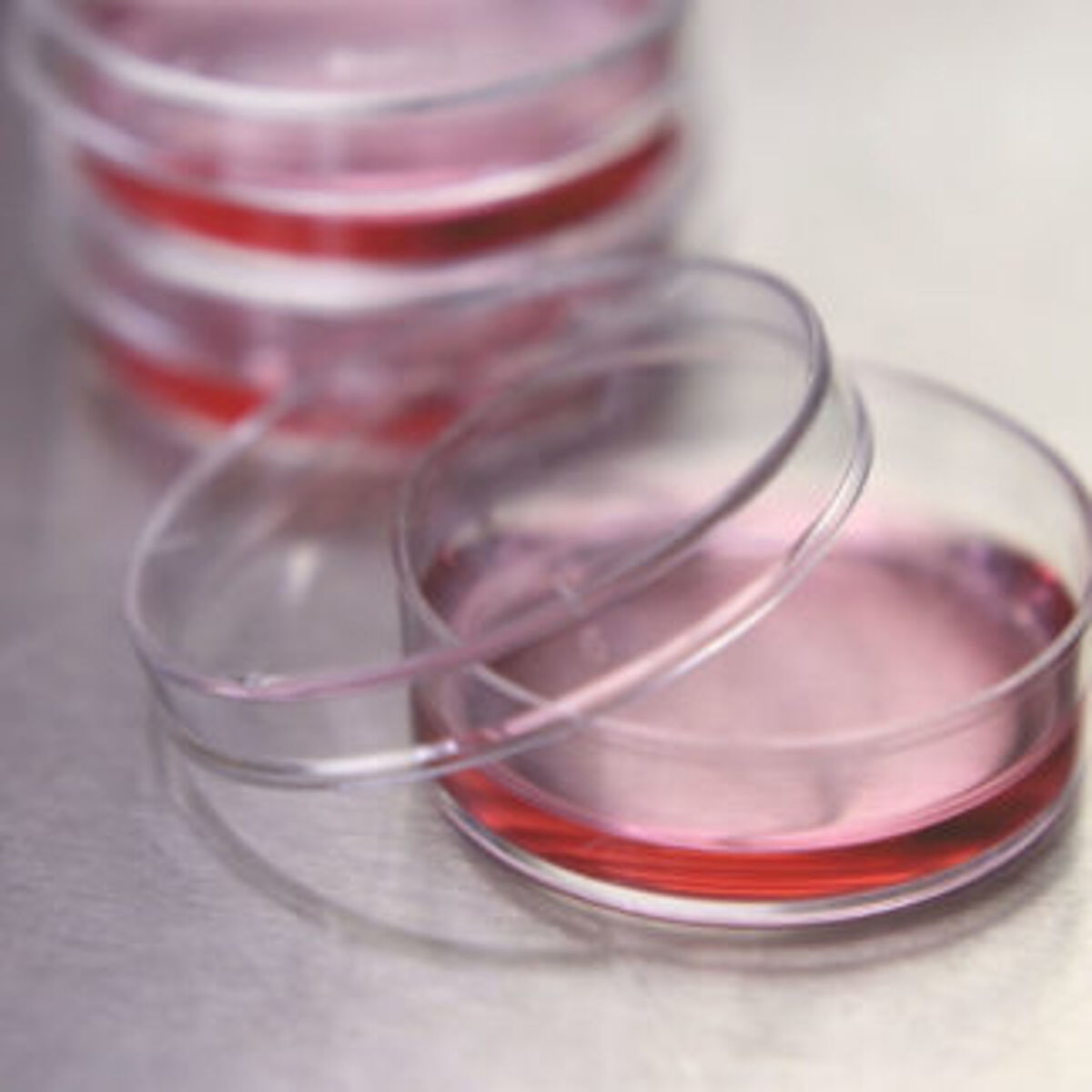
2D cell-based assays typically involve growing an immortalized cell line such as HeLa or human embryonic kidney (HEK) cells in microplate wells to form a monolayer. The cells are then treated with small molecule drug candidates and/or other entities (e.g., growth factors, cytokines) and specific responses (e.g., protein expression, cleavage, phosphorylation) are measured using techniques like ELISA, immunocytochemistry, and western blotting. Advantages of 2D cell-based assays are that they are relatively quick and easy to develop, can be scaled up for higher throughput, and often have low associated costs. However, because 2D cell-based assays do not accurately replicate conditions in vivo, the data that they generate may not translate through to a clinical setting.
Reasons for developing 3D cell models
3D cell models evolved to bridge the gap between in vitro and in vivo environments by more closely recapitulating natural conditions than traditional 2D cultures. They achieve this by allowing cells to grow and interact with one another in a three-dimensional space, often through the use of a matrix. Not only do 3D cell models provide better predictivity of in vivo responses, potentially lowering attrition rates for drug discovery, but they also serve to minimize animal use for scientific research. Since the passing of the FDA Modernization Act 2.0 in December 2022, there has been a heightened drive toward implementing 3R (reduction, refinement, replacement) practices, such as through replacing animal testing with alternative technologies1,2.
Types of 3D model systems
Because 3D cell models can vary in terms of accessibility and ease of use, it is important to understand the different options that are available. These include the following:
- Spheroids are formed by allowing one or more cell types to self-aggregate in low-attachment cultureware or propagate within a matrix. They are commonly derived from established tumor cell lines or primary tumor cells, enabling researchers to replicate the tumor microenvironment. However, spheroids may also be generated from other cell sources. For example, human gingiva-derived progenitor cells have been co-cultured with human umbilical vein endothelial cells (HUVEC) to create spheroids for studying angiogenesis3.
- Organoids are derived from embryonic stem cells, induced pluripotent stem cells, or adult stem cells that have been harvested from primary tissues. Adding different cocktails of cytokines and growth factors to the culture medium induces differentiation into miniaturized versions of organs including the lung, kidney, and pancreas. Depending on the type of model that is generated, organoids serve different purposes. For example, liver organoids can be used for assessing the risk of hepatotoxicity, which is estimated to be responsible for around 22% of clinical trial failures4.
- Microfluidic-based 3D cell culture systems are designed to control cell microenvironments, including fluid flow and gradients of nutrients and oxygen, so that tissue-specific functions are maintained in vitro. They encompass organ-on-a-chip platforms, which allow for modeling cellular interactions within specific organs, and more sophisticated body-on-a-chip systems consisting of multiple organoid types5. Recent advances within the field include the development of pumpless fluid recirculation and the introduction of biocompatible thermoplastic materials, both of which are intended to minimize drug loss due to surface adsorption6.
- 3D bioprinting, which can be broadly categorized as extrusion-, droplet-, or laser-based, uses computer-aided processes for precise layer-by-layer assembly of biomaterials. Compared with methods that allow the cells to grow randomly in culture, 3D bioprinting gives researchers tight control over cellular orientation. This can, for example, be important when modeling the intestinal mucosa, which is known to be a critical site for absorption, distribution, metabolism, and excretion (ADME) studies in drug development7.
Challenges for 3D cell culture
Working with 3D model systems presents several inherent challenges. First, the higher density of 3D structures compared with cell monolayers makes imaging more difficult, although this can be addressed through paraffin-embedding and immunohistochemical staining or by performing optical clearing8. Second, assay steps involving the transfer of liquids may require more optimization to ensure that the growing cells are not perturbed. Other issues center around the cost of 3D cell culture compared with traditional 2D cell-based assays, although demand for 3D technology looks set to drive prices down.
How can we help?
LubioScience represents some of the most trusted brands in research and works closely with our partners to offer high-quality products for both 2D and 3D cell culture. Contact us today to discuss how we can support your project.
References
- Han (2023) Artificial Organs 47(3):449-450
- NC3Rs (2023) "The 3 Rs,"
- Shanbhag et al. (2021) Frontiers in Bioengineering and Biotechnology 26:9:739225
- Zhang et al. (2023) Journal of Hepatology 78(5):998-1006
- Ma et al. (2021) Trends in Pharmacological Sciences 42(2):119-133
- Sung et al. (2018) Analytical Chemistry 91(1):330-351
- Madden et al. (2018) iScience 2:156-167
- Gonzalez et al. (2021) Journal of Visualized Experiments 27:169
Supplier
ACROBiosystems
ACROBiosystems is a leading manufacturer of recombinant proteins and other critical reagents to support the development of target therapeutics, vaccines, and diagnostics.
MCE - MedChemExpress
MedChemExpress offers high quality small molecules, reference compounds, APIs & natural compounds and inhibitor libraries. Supreme quality and rigorous QC testing by HNMR, LC-MS and HPLC - stability testing. Most compounds are tested for activity.
MBL International Corporation (MBLI)
MBL International Corporation - immunology, immuno-oncology and autoimmune diseases. MHC tetramers & monomers, antibodies, in vivo grade antibody controls.