Phage display was first reported in 1985 as a method for presenting polypeptides on the surface of filamentous bacteriophages1. It has since been harnessed for antibody discovery, where it can streamline the production of high affinity, antigen-specific antibodies for research and clinical applications.
Development of phage display technology
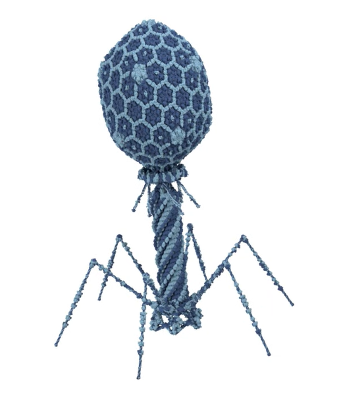
The earliest demonstration of phage display technology involved inserting foreign DNA fragments into the genome of the E. coli filamentous bacteriophage M13 such that the encoded peptide was expressed in fusion with a coat protein at the phage surface1. Critically, this provided a direct link between the phage phenotype and its encapsulated genotype, allowing for phage expressing the foreign peptide to be enriched with a peptide-specific antibody and subsequently cloned.
Just five years later, in 1990, the methodology was adapted to express antibody single-chain variable fragments (scFvs), enabling the selection of antigen-specific phage with high target affinities2. Phage display for antibody discovery has since evolved rapidly. It now incorporates the use of other E. coli bacteriophage systems (including f1, fd, T4, T7, and λ), encompasses multiple types of antibody fragments (including VH, VHH, and Fab), and has led to the construction of numerous antibody-displaying phage libraries, some containing more than 1010 phage clones.
Phage display workflow for antibody discovery
A typical phage display workflow is based on biopanning, an affinity selection technique used for enriching a desired biomolecule. In an antibody discovery setting, the target antigen is immobilized on a solid support such as a microtiter plate or a PVDF membrane, then the phage display library is added and allowed to bind. Next, any unbound phage are washed away and the bound phage are eluted for amplification in bacteria. This cycle is repeated multiple times with reduced concentrations of the immobilized antigen to select for high affinity, antigen-specific antibodies. The antibody binding activity is then assessed using ELISA, or with newer methods such as fluorometric microvolume assay technology (FMAT)3, and favorable candidates are progressed for further development.
Antibody library production
- Naïve antibody libraries are produced using V genes from non-immunized donors, which are usually derived from the IgM mRNA of donor B cells. While naïve antibody libraries are highly diverse, the antibodies that they contain often have relatively low affinities due to the fact that in vivo affinity maturation has not taken place.
- Immune antibody libraries are constructed using V genes from immunized donors or post-infection individuals, which are commonly derived from the IgG mRNA repertoire. While immune antibody libraries have less diversity than naïve antibody libraries due to being biased toward a particular antigen, this is counterbalanced by higher affinities that allow for producing smaller libraries compared to the naïve approach.
- Semi-synthetic antibody libraries are developed by introducing changes into one or more of the antibody complementary determining regions (CDRs), either through oligonucleotide directed mutagenesis or by mixing synthetic and natural sequences. Advantages of the semi-synthetic approach are that it yields a diverse set of binders with high affinities and provides opportunities to use antibody scaffolds with highly stable properties.
- Synthetic antibody libraries are generated using artificially designed and synthesized DNA oligonucleotides, which ensures complete control over the library content. Although the antibody repertoire is not shaped by natural immune maturation, the ability to introduce diversity outside of the scope of these processes allows for producing specificities that might not otherwise be attainable. For example, synthetic libraries can yield antibodies targeting toxic antigens, such as the toxic components of snake, scorpion, spider, and bee venom4. Additional advantages of the fully synthetic route are that the antibody sequences can be codon-optimized to provide high levels of in vitro expression, and there is no requirement for animal use.
Antibody phage display applications
Antibody phage display has almost limitless utility. Not only does it provide a means of producing research use only (RUO) antibodies for challenging targets, but it also has vast therapeutic potential. A recent Frontiers in Immunology publication reports that more than 70 phage-derived monoclonal antibodies have entered clinical studies, of which 14 have been approved for treating conditions that include rheumatoid arthritis, breast cancer, and neovascular age-related macular degeneration5.
LubioScience represents some of the most trusted brands in research and works closely with our partners to support antibody phage display projects. Contact us today to discuss how we can advance your studies.